Vision Technology in Flower Mound
Discover state-of-the-art vision technology at PersonalEyes Vision Care in Flower Mound. Dr. Patel and our team use advanced tools like Neurolens, Topcon Maestro2, Topcon Myah, and iCare Tonometer to provide exceptional eye care.
Experience cutting-edge vision care technology tailored to your needs.
State-Of-The-Art Equipment
At PersonalEyes Vision Care, we leverage cutting-edge vision technology in Flower Mound to deliver exceptional eye care. Our advanced equipment, from Neurolens to Topcon Maestro 2, ensures precise diagnoses and personalized treatments, enhancing your visual health and comfort.
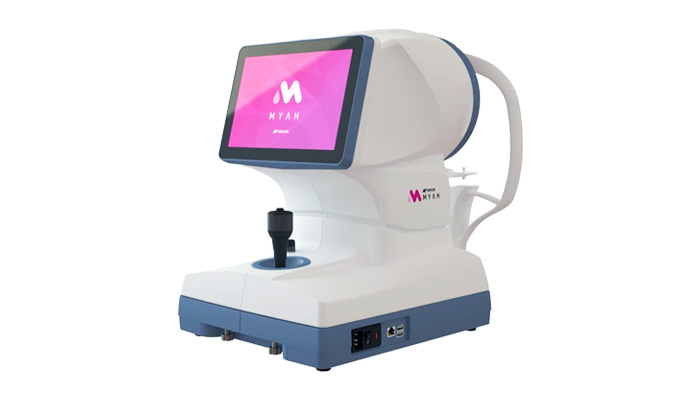
Topcon Myah - Axial Length Device
Topcon Myah is a cutting-edge device used to measure the axial length of the eye, critical for managing myopia (nearsightedness). We recommend axial length measurements every 3-6 months for children with myopia to track progression. This vision technology in Flower Mound helps prevent serious conditions like glaucoma and retinal detachment in the kids future. Early intervention is crucial and by monitoring axial length, Dr. Patel can recommend or adjust treatments like MiSight contact lenses, Johnson & Johnson's Abiliti Orthokeratology lenses, or atropine eye drops to slow myopia progression and reduce future risks.
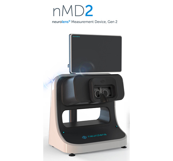
Neurolens
Neurolens is a revolutionary vision technology in Flower Mound that uses contoured prism lenses to correct eye misalignments and reduce symptoms such as headaches, neck pain, and eye strain associated with digital device use. These lenses target the muscles controlling eye alignment, bringing the eyes into a relaxed position, reducing strain. For patients with mild to severe symptoms, Neurolens can significantly improve quality of life, enabling comfortable digital device use. With growing reliance on digital devices, Neurolens is a vital tool for optometrists at PersonalEyes Vision Care.
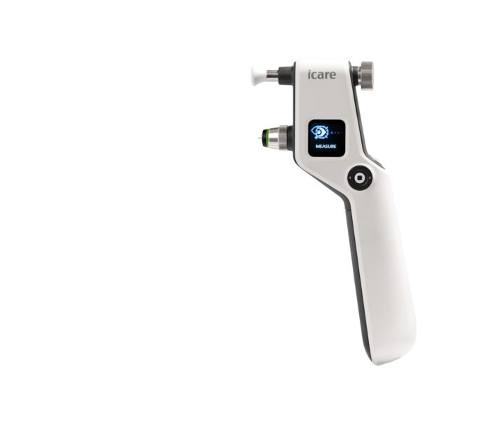
Pressure Check - iCare Tonometer
At PersonalEyes, we use the iCare Tonometer for convenient and accurate intraocular pressure evaluation, eliminating the need for the traditional "puff of air" test. This vision technology in Flower Mound aids in diagnosing and managing conditions like glaucoma. The painless, quick method provides instant and accurate pressure readings, minimally noticeable to patients, ensuring a comfortable experience.
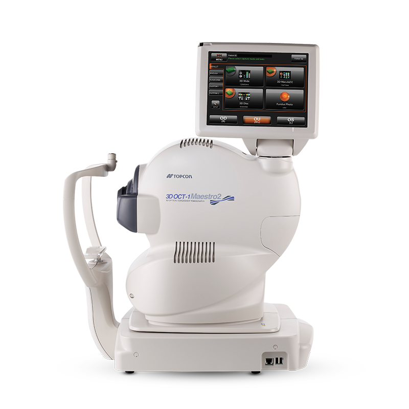
Digital Retinal Photography / Optical Coherence Tomography (OCT)
Topcon Maestro 2 provides comprehensive eye health assessments with 2-D retinal images and 3-D optical coherence tomography (OCT) scans. This vision technology in Flower Mound captures high-resolution images of the retina, essential for yearly monitoring to detect changes indicating conditions like macular degeneration, glaucoma, diabetic retinopathy, sleep apnea and many more. OCT scans reveal subtle retinal thickness changes, aiding early diagnosis. Dr. Patel notes, "Vision and health are distinct; either can affect you independently." Retinal exams and image comparisons over time help detect systemic issues like diabetes and high blood pressure.
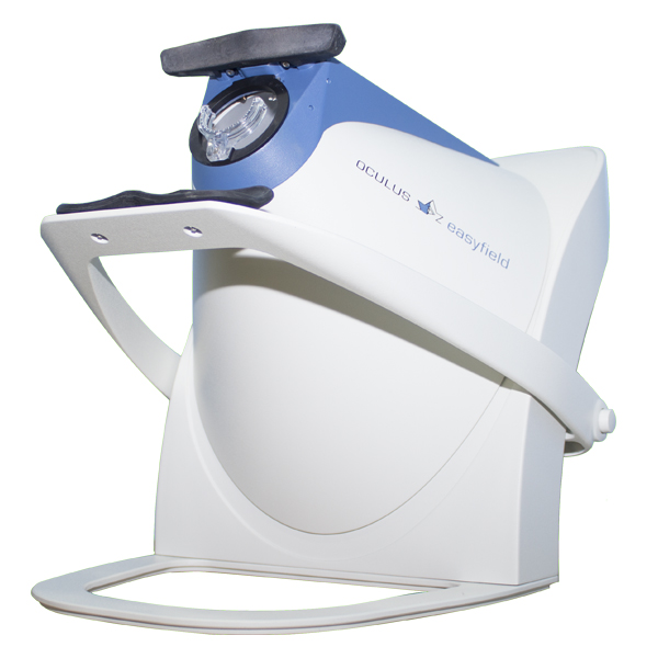
Oculus Visual Fields Test
The Oculus Visual Fields instrument assesses functional integrity by detecting small areas of missing vision. This vision technology in Flower Mound identifies defects not noticeable early due to overlapping vision in both eyes. Conditions like glaucoma, multiple sclerosis, stroke, sleep apnea, and pituitary tumors may cause visual field losses. Early detection of subtle losses is critical for timely diagnosis and treatment.
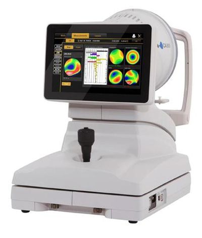
Corneal Topography / Photography / Meibomianography / Pupillometry
The TopCon CA-800 offers advanced vision technology in Flower Mound.
These tools enhance precision in eye care.
Key Scenarios for Our Vision Technology
Below are scenarios where our vision technology in Flower Mound is vital for patient care:
- When fitting rigid gas permeable lenses, Anterior Segment Photography with fluorescein ensures precise customization.
- Patients with droopy eyelids need proof of "medically necessary" for insurance coverage of blepharoplasty surgery.
- Corneal Topography Scans diagnose conditions like Keratoconus, Pellucid Marginal Degeneration, or Corneal Ectasia for better visual outcomes. Click Here To Learn More About Keratoconus!
- Pupillometry is critical when pupils appear asymmetric, aiding further investigation like cranial MRI or chest X-Ray.
Reading time: 5 minutes
Last updated: July 8, 2025